Recent publications

Graphical abstract showing a mouse-specific aorta model with simulation panels linking high endothelial cell activation potential to thoracic aortic aneurysm progression Graphical abstract showing a mouse-specific aorta model with simulation panels linking high endothelial cell activation potential to thoracic aortic aneurysm progression

Dwivedi et al., Comput Biol Med, 2026

Comput Biol Med. 2026 April:205:111594 doi:10.1016/j.compbiomed.2026.111594

Graph of aortic diameter over time comparing EGCG plus elastase treatment with untreated vessels, with fluorescent images of elastic fibers Graph of aortic diameter over time comparing EGCG plus elastase treatment with untreated vessels, with fluorescent images of elastic fibers

Castro, Chen et al., JMBBM, 2026

J Mech Behav Biomed Mater. 2026 Apr:176:107340. doi: 10.1016/j/jmbbm.2026.107340

Side-by-side fluorescence microscopy images of aortic tissue from two Eln/Cre mouse genotypes with blue-stained nuclei Side-by-side fluorescence microscopy images of aortic tissue from two Eln/Cre mouse genotypes with blue-stained nuclei

Ruddiman et al., Hypertension, 2025

Hypertension. 2025 Dec;82(122):2112-2124. doi:10.1161/HYPERTENSIONAHA.124.24372.

Grayscale and color-mapped polarized imaging panels of two tissue samples showing degree and angle of polarization Grayscale and color-mapped polarized imaging panels of two tissue samples showing degree and angle of polarization

Pavey et al., J Biomech Eng, 2025

J Biomech Eng. 2025 Nov 1:147(11):111010. doi: 10.1115/1.4069533.

Stress–stretch surface plots comparing circumferential and axial aortic mechanics in wild-type and mutant mice Stress–stretch surface plots comparing circumferential and axial aortic mechanics in wild-type and mutant mice

Wu et al., JMBBM, 2025

J Mech Behav Biomed Mater. 2025 Oct:170:10705. doi: 10.1016/j.jmbbm.2025.107105.
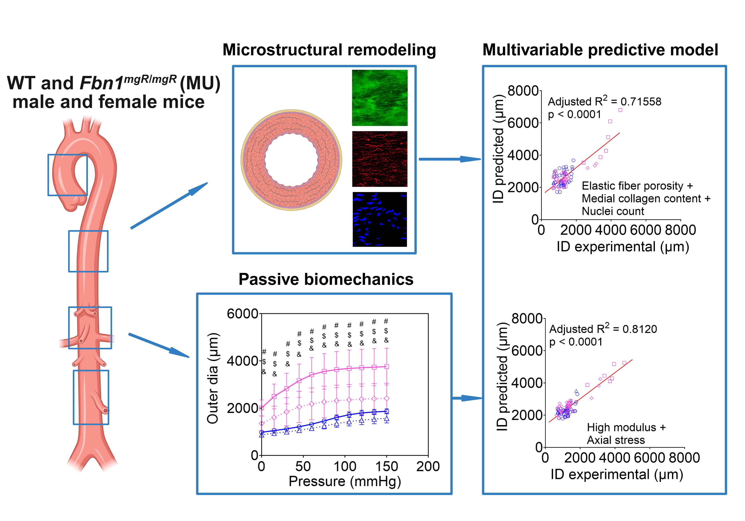
Diagram linking microstructural remodeling and passive biomechanics of wild-type and Marfan mouse aortas to a multivariable predictive model Diagram linking microstructural remodeling and passive biomechanics of wild-type and Marfan mouse aortas to a multivariable predictive model

Dwivedi et al., Acta Biomat, 2025

Acta Biomater. 2025 May 23:S1742-7061(25)00382-4. doi: 10.1016/j.actbio.2025.05.056. Online ahead of print.

Illustration of a thoracic aortic aneurysm and aortic wall cross-section annotated with imaging agents and therapeutic molecules for diagnosis and treatment Illustration of a thoracic aortic aneurysm and aortic wall cross-section annotated with imaging agents and therapeutic molecules for diagnosis and treatment

Kailash et al., AJP Heart Circ, 2025

Am J Physiol Heart Circ Physiol. 2025 May 1;328(5):H1113-H1129. doi: 10.1152/ajpheart.00886.2024. Epub Apr 7.

Illustration of an aortic wall cross-section with particles moving toward smooth muscle cells, depicting transmural transport Illustration of an aortic wall cross-section with particles moving toward smooth muscle cells, depicting transmural transport

Kailash et al., BMMB, 2024

Biomech Model Mechanobiol. 2024 Oct 29. doi: 10.1007/s10327-024-01897-5.

Diagram of the Marfan mouse aorta with graphs comparing dilation and stiffening/ECM remodeling over age in males and females Diagram of the Marfan mouse aorta with graphs comparing dilation and stiffening/ECM remodeling over age in males and females

Dwivedi et al., AJP Heart Circ, 2024

Am J Physiol Heart Circ Physiol. 2024 Oct 1;327(4):H1037-H1051. doi: 10.1152/ajpheart.00391.2024.

Histology panels of male and female Eln mouse aortas with graphs quantifying medial collagen and other tissue components Histology panels of male and female Eln mouse aortas with graphs quantifying medial collagen and other tissue components

Kailash et al., J Biomech Eng, 2024

J Biomech Eng. 2024 Mar 1;146(5):051001.doi: 10.115/1.4063272

Journal cover figure relating transmural mass transport in the aortic wall to aneurysm size and elastic fiber fragmentation Journal cover figure relating transmural mass transport in the aortic wall to aneurysm size and elastic fiber fragmentation

Crandall et al., AJP Heart Circ, 2023

Am J Physiol Heart Circ Physiol. 2023 Jul 1;325(1):H113-H124. doi: 10.1152/ajpheart.00036.2023. Epub 2023 Jun 2.

Illustration of myoendothelial junctions in the vessel wall comparing regions with and without PS signaling Illustration of myoendothelial junctions in the vessel wall comparing regions with and without PS signaling

Ruddiman et al., JCI Insight, 2023

JCI Insight. 2023 May 8;8(9):e165715. doi: 10.1172/jci.insight.165715.

Fluorescence images of elastic fibers in the ascending and descending thoracic aorta of wild-type and Eln mutant mice Fluorescence images of elastic fibers in the ascending and descending thoracic aorta of wild-type and Eln mutant mice

Brengle et al., Matrix Biol, 2023

Matrix Biol. 2023 Feb 9;S0945-053X(23)0013. doi:10.1016/j.matbio.2023.02.003

Diagram of ECM turnover by enzymes and macrophages in the juvenile mouse aorta, contrasting a normal aortic wall with progressive ECM degradation and TAAD Diagram of ECM turnover by enzymes and macrophages in the juvenile mouse aorta, contrasting a normal aortic wall with progressive ECM degradation and TAAD

Wagenseil, Biomat Biosys, 2023

Biomat Biosys. 2023, Mar;9:100072. doi:10.1016/j.bbbiosys.2023.100072.